| Portosystemic Shunt CT angiography revealed multiple tortuous vessels near the left kidney,esophageal varices,and signs of primary liver disease. |
|
| Thoracic Duct Imaging Used to evaluate congenital/acquired limb edema, chylothorax causes,surgical outcomes,and tumor-related sentinel lymph nodes |
|
| Tympanic Empyema CT imaging showed purulent fluid accumulation in the tympanic cavity with surrounding inflammation,correlating with head-shaking and ear discharge. |
| Part | Case |
| Head&Neck | Cerebral Edema,Brain Hemorrhage,Sinusitis,Otitis,Mastoid Effusion,Nasopharyngeal Stenosis,Tracheal Narrowing,etc. |
| Dental | Periodontitis,Periapical Abscess,Alveolar Bone Atrophy,Oral Cyst,Tooth Root Resorption,etc. |
| Thoracic | Pulmonary Edema,Pulmonary Nodules,Interstitial Pneumonia,Pulmonary Inflammation,etc. |
| Abdomen | Abdominal Wall Inflammation,Peritoneal Fluid Accumulation,Urography,Renal Imaging,Gallstones,Pancreatitis,Kidney Hardening. |
| Vascular | Abdominal Three-Phase Angiography,Peripheral Artery Thrombosis,Vascular Stenosis,etc. |
| Spine | Spinal Canal Imaging,Spinal Protrusion,Vertebral Imaging,Spondylosis,Thoracic Spine Fracture,etc. |
| Tumors | Thyroid Tumors,Nasal Tumors,Ovarian Tumors,Renal Tumors,Adrenal Tumors,etc. |
| Others | Arthritis,Thoracic Duct Imaging,Peritoneal Calcification,etc. |
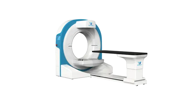
| Gantry | Structure | Ring-shaped |
| Aperture | 31.5″(80cm) | |
| Nominal Power | 15kW | |
| Input Power | 220Vac±10% | |
| X-ray Tube Heat Capacity | 150kJ | |
| Table | Horizontal Movement | 28.3″(720mm) |
| Vertical Movement | 11″(280mm) | |
| Scan Parameters | CT Scan Slices | 600-1000 |
| CT Slice Thickness | 0.2-0.72mm | |
| CT Scan Time | 4.5s | |
| Max.FOV(L x W) | 28.3″×16.5″ (72cm×42cm) |