
I’m going to be direct about the Vetease 3 because this is a significant capital investment and nobody needs a sales pitch when they’re evaluating a six-figure imaging system. The question every veterinary practice owner needs to answer before looking at CT is: how many cases per month are you referring out to a university hospital or a specialty imaging center for advanced diagnostics? If the answer is more than five, you’re probably losing revenue, losing client relationships, and losing treatment windows that depend on fast imaging turnaround. The Vetease 3 exists to bring CT capability in-house at a price point that makes that math work for mid-size and growing veterinary hospitals.
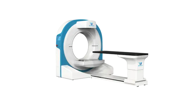
The Vetease 3 is a ring-shaped gantry CT scanner purpose-built for veterinary use. The aperture is eighty centimeters — thirty-one and a half inches — which accommodates large-breed dogs in sternal or dorsal recumbency without the positioning compromises that happen when you try to squeeze a Great Dane into a human CT scanner. The gantry design is fixed-ring, not a rotating C-arm, which means the imaging geometry stays consistent across the full scan field. Nominal power is fifteen kilowatts with a tube heat capacity of one hundred fifty kilojoules, so you can run consecutive scans without waiting for the tube to cool between patients — a genuine workflow consideration in a busy practice.
Imaging specs: six hundred to one thousand slices per scan series, with selectable slice thickness from zero point two to zero point seven two millimeters. At the thinnest slice setting, the resolution is fine enough to visualize small vessel anatomy, dental root structures, and early-stage tumor margins that would be invisible on radiographs. Scan time is four point five seconds for a full rotation — fast enough that most animals can be scanned under sedation rather than general anesthesia, which significantly reduces the risk profile and recovery time. The maximum field of view is seventy-two by forty-two centimeters, covering the full body cross-section of all but the most massive giant-breed dogs.
Clinical applications span the full veterinary imaging spectrum: head and neck (cerebral edema, sinusitis, otitis, nasopharyngeal stenosis, tracheal narrowing), thoracic (pulmonary edema, nodules, interstitial patterns), abdominal (peritoneal fluid, urography, renal imaging, gallstones, pancreatitis), vascular (three-phase angiography, thrombosis, stenosis), spinal (canal imaging, disc protrusion, vertebral assessment, spondylosis), dental (periodontitis, periapical abscess, alveolar bone atrophy, oral cysts, root resorption), and tumor imaging across all anatomical regions. The system also supports thoracic duct imaging for congenital and acquired lymphatic conditions — a specialized but growing area of veterinary surgical planning.
Input power is standard two hundred twenty volts AC, which means it runs on the same electrical infrastructure as most imaging suites — no three-phase industrial power required. This matters when you’re retrofitting an existing clinic rather than building new construction. The table has seven hundred twenty millimeters of horizontal travel and two hundred eighty millimeters of vertical movement, so positioning is flexible without excessive manual repositioning between series. Installation includes shielding assessment and room planning — I’ll be honest, you need a dedicated room with appropriate lead lining, and that’s a cost you should factor into your total budget from the start. If you’re at the evaluation stage, send me your monthly referral volume and the primary clinical applications you’re targeting. I’ll help you run the numbers and decide whether an in-house CT makes financial sense for your practice size.